
New cardiological research in The „New Life” Clinic
Transesophageal echocardiography is a vital research approach in cardiology that enables the precise diagnosis of abnormal alterations in the heart. (TEE) is conducted using ultrasound, therefore the patient is not exposed to radiation.
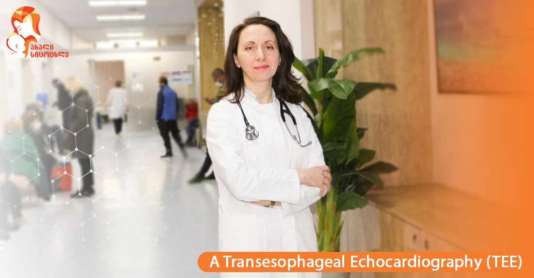
Inga Zhvania, a cardiologist at the National Center of Surgery’s “New Life” Clinic, explains the research process in greater detail:
-Transesophageal echocardiography is a non-invasive ultrasonographic examination of the heart, a so-called ultrasonic transmitter, a flexible probe is placed at heart level into the esophagus. Because of the close proximity of the esophagus to the heart, this method produces a more accurate image of the heart than conventional, transthoracic echocardiography, because the chest muscles, ribs, subcutaneous fat, or lung tissue thwart ultrasound from reaching the heart and, in some cases, aggravate visualization of heart structures.
-When, and in what cases is transesophageal echocardiography performed?
-Traditional transthoracic echocardiographic examinations provide comprehensive information for assessing the structure and function of the heart, but it is occasionally required to view the structure of the heart with specific accuracy. At this point, the TEE is recommended. For example, we can obtain a full-fledged image of the left atrial ear appendage, which is prone to the formation of thrombotic masses, particularly during arrhythmias. As a result, the probability of forming a thrombus mass and having a stroke increases. As a result, this test is required prior to performing cardioversion, a treatment to restore cardiac rhythm. Furthermore, it is regarded as the gold standard for diagnosing heart valves. When endocarditis or the presence of a cardiac tumor is suspected, it is distinguished by its information quality. Used to evaluate acquired and congenital thoracic aortic and wall abnormalities. TEE can also be performed during open-heart surgery and intracardiac transcatheter operations.
-How is the (TEE) research procedure conducted?
-TEE, like a gastroscopic examination, is done on an empty stomach. Food and fluids should not have been consumed by the patient for at least six hours. The procedure takes 20-30 minutes and is not uncomfortable. The doctor inserts the probe to the proper level in the esophagus, and we capture a real-time image of the heart on the monitor. The examination may be performed under local anesthetic and sedation if necessary.
-What are the study's potential risks?
-During the examination, the patient's vital signs are continuously monitored. Although unlikely, teeth, esophagus, and gastric mucosa may have been harmed. Anesthetics may produce an allergic reaction, shortness of breath, or arrhythmia. However, the chance of life-threatening complications is less than 0.1 percent in general.
-What steps are performed following the examination?
-Following the procedure, the patient's vital indicators are evaluated, and the patient is observed until the sedative medicine wears off. The patient should refrain from eating or drinking for 2-3 hours, leave the clinic with a companion, and not drive for 24 hours.
The „New Life“ Clinic wishes you health!