Computed tomography is a high-technology method of study in diagnostic radiology.
In the interview, you will be informed about the following:
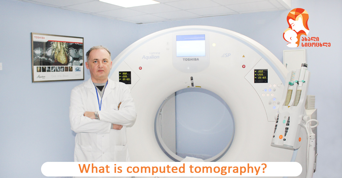
The clinic “New Life” of National Center of Surgery is equipped with ultramodern, 160-slice tomography scanner Toshiba Aquilion Lightning SP that has all the necessary latest systems of study.
One of the main advantages of the device is 160 slices that significantly reduce scanning time and covers more body range in one rotation.
Study is conducted quickly, effectively and safely.
The clinic’s tomography scanner is distinguished by diagnosing measures, images, and information capability.
The clinic offers the study to bariatric (heavy weight) patients who weigh under 255 kg.
What is computed tomography (CT)?
It’s possible to get a layered 3D image of organs and tissues with it using x-ray beams.
This is a full-body image.
The radiologist sees in detail the ongoing changes in the body and, reaches an appropriate diagnosis after identifying various types of pathologies
The study is conducted on a scheduled, as well as urgent (emergency) basis.
Various surgical manipulations are conducted with CT control, it’s necessary for biopsy and radiation therapy.
Radiologist of the clinic “New Life” of National Center of Surgery, Giorgi Pipia discusses the topic.
– Study of which organs is computed tomography used for?
– CT is used for the detection and monitoring of numerous diseases and pathologies.
It’s conducted to diagnose and monitor chronic diseases, in particular: cancer pathologies, systemic diseases, to detect and monitor damages to the brain, lung, liver and biliary system, spleen, urinary system, adrenal glands and digestive tract, skeletal system.
It’s necessary for diagnosing such acute damages as:
- Traumas, in case of which this method is practically indispensable because it enables the assessment of the damage and severity of various organs in the short time;
- Brain stroke (differentiation of ischemic and hemorrhagic strokes, damage detection at the early stage with CT perfusion and CT angiography, identifying/planning prognosis and treatment tactics);
- Spinal injury and so on.
This study method also enables diagnosing and post-operative monitoring of following angiological (vascular) pathologies:
- Aneurysm;
- Malformation;
- Identifying the grade of stenosis and occlusion.
In our clinic, we conduct practically all types of studies for 24 hours.
– What does it mean to prepare the patient for computed tomography?
– Preparation for the study doesn’t require any special rules. Albeit, several issues should be considered, in particular, abdominal and pelvic computed tomography isn’t informative if the patient took Barium for several days. In this case, we provide respective advice-recommendations to the patient and then conduct the study.
In the case of diabetes, a patient might need to stop consuming certain medications a day before and on the day of the procedure.
An allergic patient must inform medical personnel about the allergy – when an allergic patient undergoes a study with intravenous contrast, allergologist, together with a radiologist, care for patient's health, and the study is carried out only after appropriate preparation.
In case of a high level of creatinine, nephrologist participates in treatment and computed tomography is ordered only after the consultation with a said specialist.
The study is contraindicated in pregnant women, nursing mothers. In this case, the study is conducted only as an emergency or vital procedure. Even in this case, it’s recommended for the procedure to proceed with particularly gentle protocol.
– If a patient's afraid of closed spaces (claustrophobia), what alternative do you offer?
– In the case of claustrophobia, we show the device in advance to the patient, the amplitude of its movement, depth, and frequency. We explain that the study process lasts from 5 to 10 minutes on average (of course, it all depends on the complexity of the study), we are always in touch with the patient using a microphone and, if necessary, we can interrupt the procedure at any time. If all these attempts fail, after consultation with the anesthesiologist we conduct the study with intravenous anesthesia.
-What does computed tomography with contrast mean?
- The study is conducted with or without contrast. Doctor-in-charge decides which method is appropriate. Moreover, the radiologist's recommendation has to be considered for better visualization and differentiation of details in the study process.
The difference between studies with and without contrast depends on the type of pathology we want to determine. Albeit, in most cases the contrast method is recommended for high accuracy.
Different tissues of the body can stop the x-ray beams (in most cases, mass and healthy tissue have similar density) and because of that image might not be comprehensive for diagnosing; intravenous contrast substance is used in such cases.
Iodine-based contrast substance enables the radiologist to conduct a full assessment of blood vessels (without the contrast vascular study is not informative) and is used to determine possible vascular obstructions, whereas, peroral contrast substance is used for diagnosing gastrointestinal tract organs.
In I/V contrast, pathologies are mostly detected in arterial or venous phases, in most cases, radiologist uses delayed phase to differentiate: for example, in between a liver hemangioma and other masses, because effect and nature of contrast enhancement (peripheral or central enhancement), time and volume of wash-out (complete wash-out, incomplete wash-out-delay) enables us to determine the type of the process we’re dealing with.
Moreover, for example, if the brain cyst is identified, I/V contrast enables us to determine if the mass is benign (meningioma) or secondary injury – MTS (ring-enhancing), or malignancy is suspected.
Final diagnosis, of course, is determined on the basis of cytomorphology, albeit primary radiological indicators help clinician plan follow-up treatment and studies.
Wish you health!