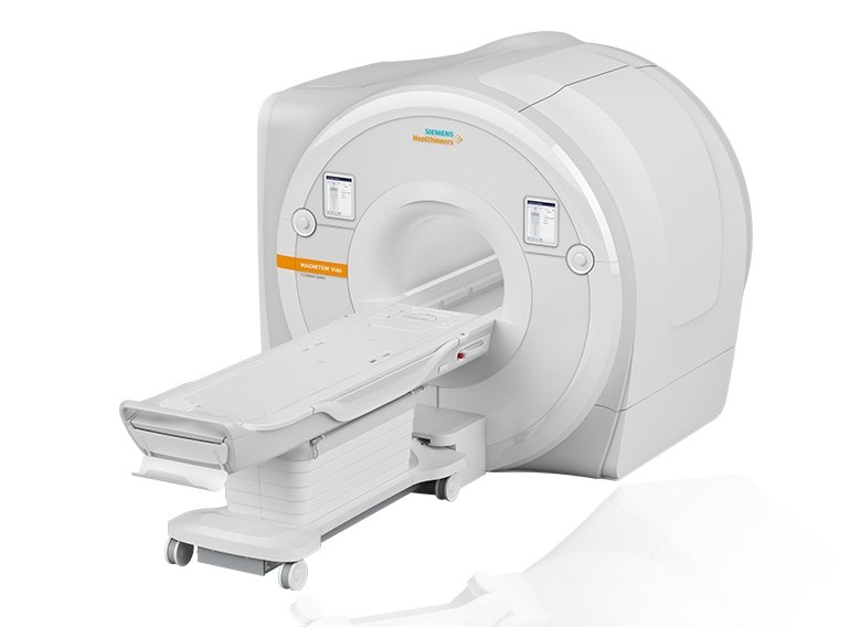
Siemens Magnetom Vida 3T, a digital artificial intelligence 3 Tesla magnetic resonance tomography manufactured in Germany, is utilized in the clinic of the National Center of Surgery’s ,,New Life“.
It should be mentioned that this examination technique is entirely safe for human health because no ionized radiation is employed, and no human tissues or internal organs are exposed to X-ray radiation during the course of the study.
MRI imaging serves an essential part in tracking the course of numerous chronic diseases, including cancer.
With its robust and cutting-edge Tim 4G design, the 3 Tesla MRI produces the best possible images, enabling radiologists to identify even the smallest anatomical structures and ensuring precise diagnosis.

One of the best MRI models is available at the National Center of Surgery's "New Life" clinic. It's a new generation digital magnetic resonance tomography with artificial intelligence, which was selected for our facility due to its many benefits and advances:
- The BioMatrix technology, which offers intelligent patient customisation, is integrated into Magnetom Vida;
- HyperBand shortens the duration of each scan. Patients who struggle to remain still will benefit most from this technology;
- Compressed Sensing is a cutting-edge imaging method that is transforming medical imaging;
- GOBrain: The GOBrain app is integrated with the Magnetom Vida 3T, enabling a thorough neurological evaluation of the brain in just five minutes;
- The goal of Quiet Suite technology is to lessen background noise while scanning. It makes use of combined software and hardware advancements;
- Motion artifacts are minimized and image quality is enhanced with free-breathing imaging;
- Native Direct MR arthrography is a feature of the Magnetom Vida 3T that enables high-quality picture acquisition without the need for additional contrast agents. It is especially useful for assessing injuries to the cartilage and tendons;
- TimTX TrueForm: this technology improves image quality and minimizes artifacts, particularly in challenging clinical scenarios involving heavier patients or particular body parts that are difficult to reach;
- Magnetic resonance spectroscopy (MRS): this technique uses an analysis of the signal produced by different metabolites to ascertain the chemical composition of tissues. It offers insightful knowledge regarding tissue metabolism. mostly utilized for tumor evaluation and neuroradiology;
- Functional MRI (fMRI): a technique for examining brain activity and brain function that measures variations in blood oxygenation;
- Cardiac MRI: This diagnostic tool makes it possible to assess the anatomy and physiology of the heart. It offers comprehensive details regarding the size, blood flow, and viability of the heart.